
 |


| Quantity: | |
|---|---|
LPH-101-2.5
Piezohannas
LPH-101-2.5
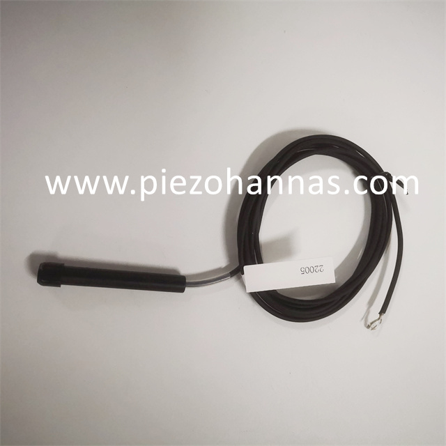
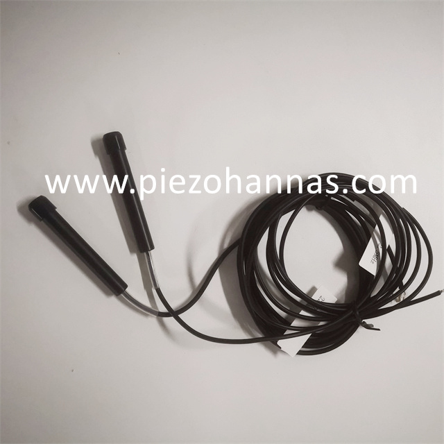
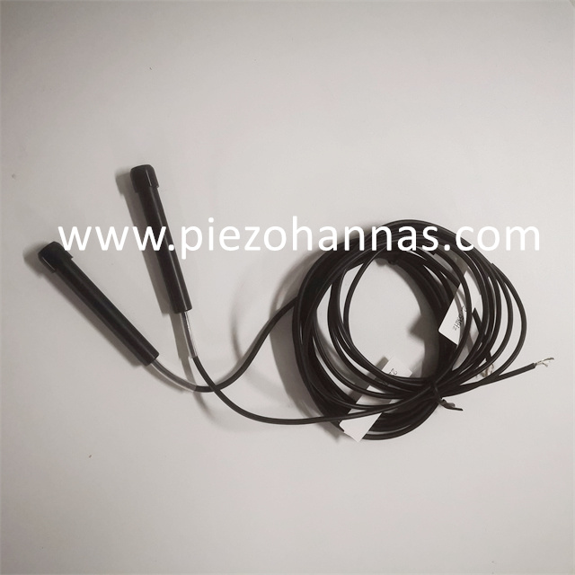
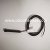
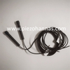
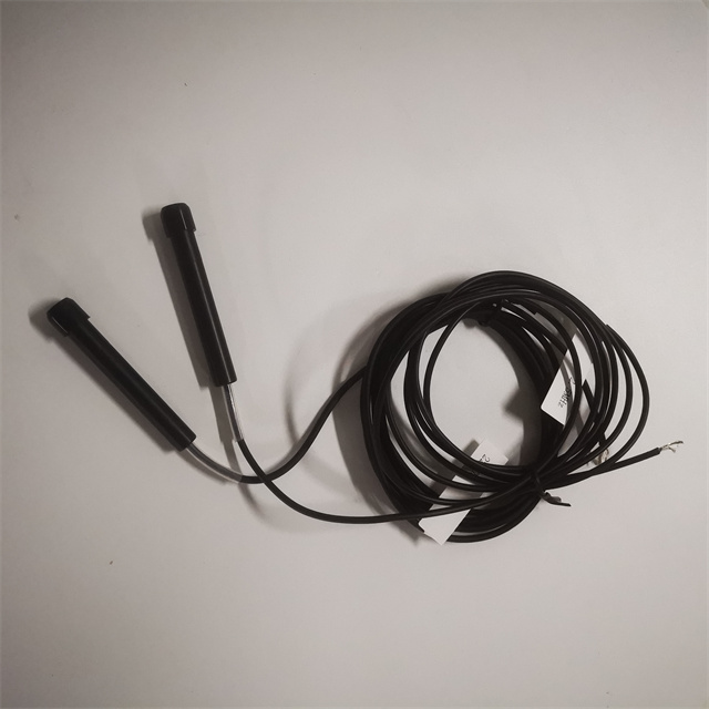
Custom 2.5Mhz A-scan Medical Ultrasound Probes Ultrasonic Sensor
Specification:
2.5Mhz A -scan Ultrasound Probes | |||||||
Pulse echo maximum response frequency fP(Mhz) | Pulse echo bandwidth△f | pulse center frequency F0(Mhz) | Pulse echo width (duration) (μs) | Relative pulse echo sensitivity level (dB) | Total resonant impedance (Ω) | Static capacitance(Pf) | Pressure resistance 4000V |
2.55 | 1.5 | 2.6 | 1.7 | -24.0 | 81 | 1170 | √ |
2.55 | 1.5 | 2.5 | 1.7 | -24.0 | 85 | 1170 | √ |
2.50 | 1.5 | 2.5 | 1.7 | -24.0 | 84 | 1160 | √ |
2.55 | 1.5 | 2.5 | 1.7 | -24.0 | 83 | 1170 | √ |
2.55 | 1.5 | 2.5 | 1.7 | -24.0 | 98 | 1220 | √ |
Working Principle:
The ultrasound A-scan works by projecting a sound wave through the eye and then measuring reflections. A reflection happens at the junction of two different densities, such as the interface between the posterior lens capsule and the vitreous. These echoes are shown as tall spikes of a high amplitude on the display, hence the name amplitude-scan, which is often abbreviated as A-scan.
A-scan ultrasound is used to take measurements for artificial lenses for cataract surgery. This quick and painless procedure can be performed in your doctor’s office.
Application Image:
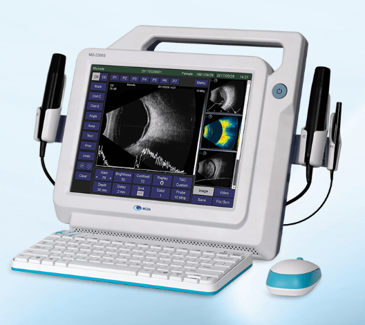
 Tech Co,.Ltd")