
 |


| Quantity: | |
|---|---|
LPH-10
Piezohannas
LPH-10
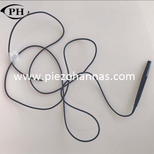
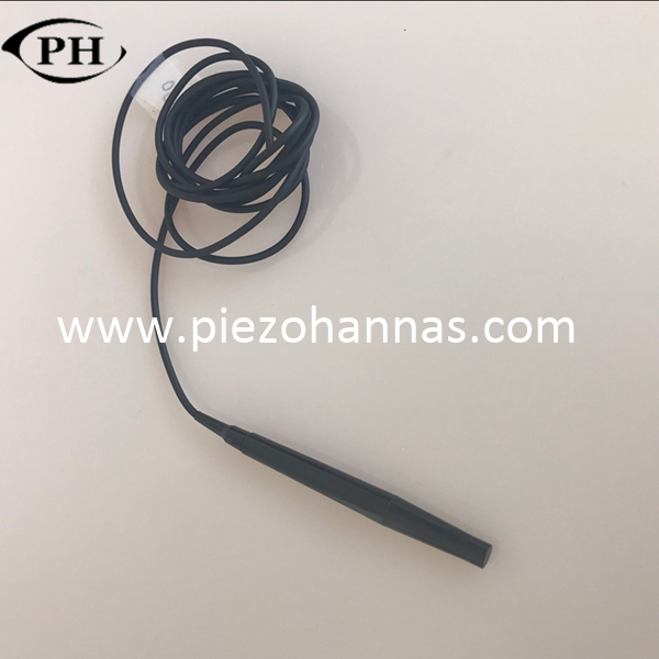
10Mhz Ultrasound A-scan Transducer Ophthalmic A-mode Ultrasonoscope for Small Animals
Specification:
Name | 10MHz medical phthalmology probe | |||
Number | LPH-10MHz | Product Standard | Enterprise Standard | |
Structural Material | Performance Index (Water) | |||
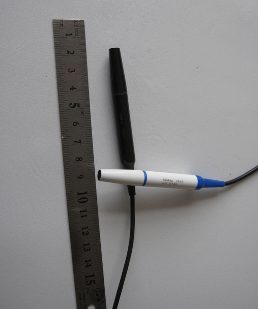
Piezoelectric Ceramics | Size | Ф4.8×0.2 mm | Resonant Frequency | 10MHz±10% |
Material | PZT-5 | Relative Bandwidth | >65% | |
Capacitance | 400pf ±10% | Pulse Width | < 300ns | |
Transceiver Sensitivity | > -35dB
| |||
Cable | Ф3mm twin-core shield cable
| |||
Shell | Size | Ф9×75 mm | ||
Material | ABS | |||
Contacting Material on the front-end and eyeball | 5010A/5010B epoxy resin encapsulating material | |||
Technical Parameters | 5M | 7.5M | 10M | 13M |
Operating frequency | 5MHz | 7.5MHz | 10MHz | 13MHz |
Relative bandwidth | >50% | >50% | >60% | >60% |
relative sensitivity | >-24dB | >-30dB | >-35dB | >-42dB |
Echo pulse width | <0.7μs | <0.55μs | <0.4μs | <0.35μs |
Application | For pets, livestock measuring fat | For pets, livestock measuring fat | For measuring distance in ophthalmology | For measuring thickness in ophthalmology |
Working Principle:
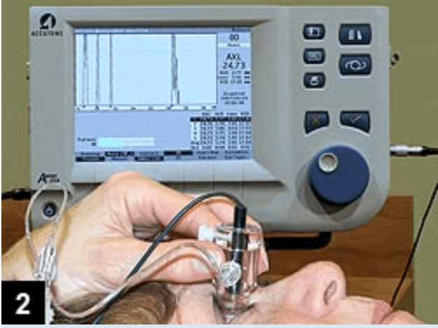
The ultrasound A-scan works by projecting a sound wave through the eye and then measuring reflections. A reflection happens at the junction of two different densities, such as the interface between the posterior lens capsule and the vitreous. These echoes are shown as tall spikes of a high amplitude on the display, hence the name amplitude-scan, which is often abbreviated as A-scan.
A-scan ultrasound is used to take measurements for artificial lenses for cataract surgery. This quick and painless procedure can be performed in your doctor’s office.
Application Image:

10Mhz Ultrasound A-scan Transducer Ophthalmic A-mode Ultrasonoscope for Small Animals
Specification:
Name | 10MHz medical phthalmology probe | |||
Number | LPH-10MHz | Product Standard | Enterprise Standard | |
Structural Material | Performance Index (Water) | |||
Piezoelectric Ceramics | Size | Ф4.8×0.2 mm | Resonant Frequency | 10MHz±10% |
Material | PZT-5 | Relative Bandwidth | >65% | |
Capacitance | 400pf ±10% | Pulse Width | < 300ns | |
Transceiver Sensitivity | > -35dB
| |||
Cable | Ф3mm twin-core shield cable
| |||
Shell | Size | Ф9×75 mm | ||
Material | ABS | |||
Contacting Material on the front-end and eyeball | 5010A/5010B epoxy resin encapsulating material | |||
Technical Parameters | 5M | 7.5M | 10M | 13M |
Operating frequency | 5MHz | 7.5MHz | 10MHz | 13MHz |
Relative bandwidth | >50% | >50% | >60% | >60% |
relative sensitivity | >-24dB | >-30dB | >-35dB | >-42dB |
Echo pulse width | <0.7μs | <0.55μs | <0.4μs | <0.35μs |
Application | For pets, livestock measuring fat | For pets, livestock measuring fat | For measuring distance in ophthalmology | For measuring thickness in ophthalmology |
Working Principle:
The ultrasound A-scan works by projecting a sound wave through the eye and then measuring reflections. A reflection happens at the junction of two different densities, such as the interface between the posterior lens capsule and the vitreous. These echoes are shown as tall spikes of a high amplitude on the display, hence the name amplitude-scan, which is often abbreviated as A-scan.
A-scan ultrasound is used to take measurements for artificial lenses for cataract surgery. This quick and painless procedure can be performed in your doctor’s office.
Application Image:
 Tech Co,.Ltd")